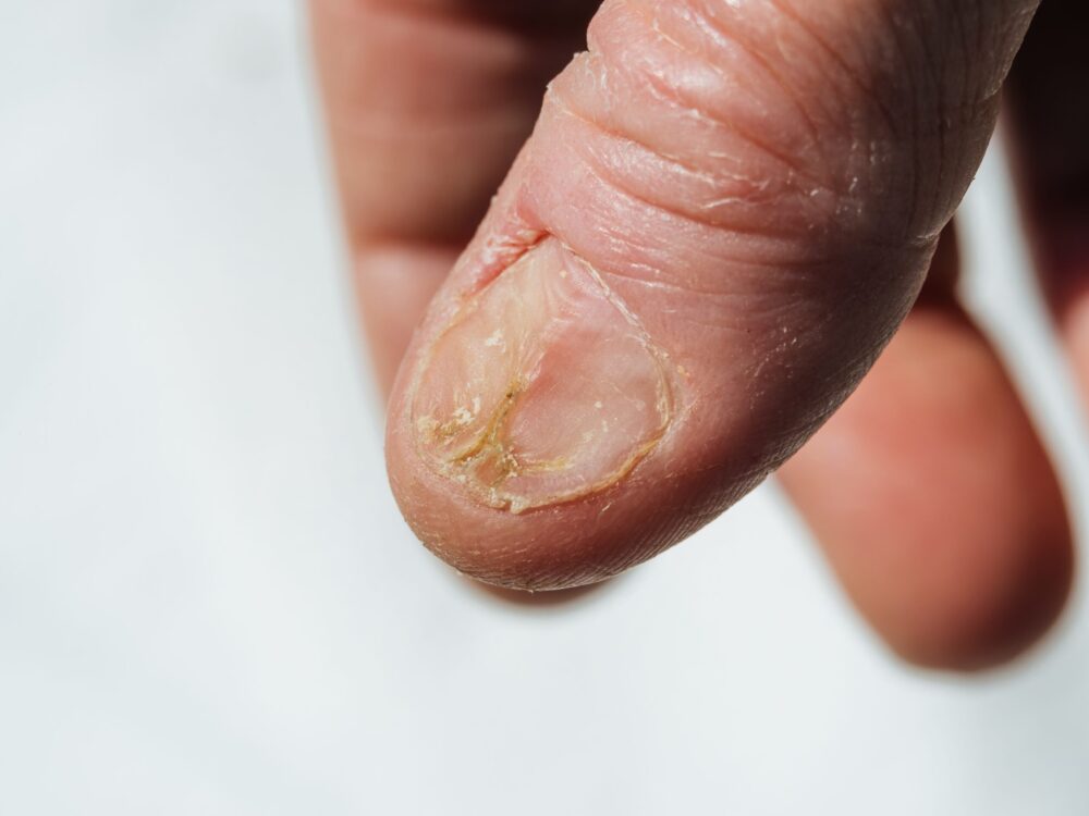
What your fingertips are quietly communicating
These surface changes are rarely the straightforward story they appear to be. A brittle edge, an unexpected discoloration, a ridge that was not there six months ago, all are easy to attribute to aging, to a new hand wash, or to the last gel manicure. They deserve a more considered read. The nail is a living structure, continuously produced by the matrix at its base and sensitive to the nutritional, circulatory, hormonal, and immunological conditions of the body that grows it.
Clinicians who examine this surface routinely look for evidence of iron deficiency, thyroid dysfunction, psoriasis, fungal infection, liver disease, cardiovascular insufficiency, and in some cases early signs of malignancy, all before these conditions have produced other obvious symptoms. Understanding what changes mean is not about diagnosing from a photograph. It is about knowing which shifts warrant a provider conversation rather than a visit to the salon.
Reading what discoloration signals
Color changes in the plate and underlying bed carry some of the richest diagnostic information available through visual examination alone. White nails, where most of the nail appears pale with a narrow band of pink at the tip, are associated with liver disease, kidney failure, and in some presentations, heart failure. Entirely pale nails can reflect anemia, where reduced red blood cell concentration diminishes the pinkish hue that healthy circulation provides to the nail bed.
A yellow appearance is most frequently caused by fungal infection, which thickens and discolors the plate from the leading edge inward. Less commonly, yellow nails appear in association with lymphatic system disorders or chronic respiratory conditions. Green-black discoloration usually indicates bacterial infection of the underlying bed, typically following trauma that allows bacteria to establish beneath the plate.
Dark streaks running vertically from the matrix to the free edge require particular attention. In people with lighter skin tones, a new or changing dark longitudinal band should be evaluated by a dermatologist, as a small proportion represent subungual melanoma, a form of skin cancer originating beneath the plate.
Structural shifts and what causes them
Small depressions scattered across the surface of the plate, known as pitting, are one of the most reliable physical signs of psoriasis. It appears in nail tissue before skin plaques develop in a significant percentage of people with the condition and can be the finding that prompts an earlier diagnosis of a systemic inflammatory disease that extends well beyond the skin.
These horizontal grooves or indentations cross the full width of the keratin plate and are known as Beau’s lines. They form when the nail matrix temporarily stops producing nail tissue, a response to physical or physiological stress including severe illness, surgery, chemotherapy, or significant nutritional deficiency. Because nails grow at a predictable rate, the position of a Beau’s line can be used to estimate when the disrupting event occurred.
Spoon-shaped curvature, where the edge curves upward rather than following the natural arc of the fingertip, are associated with iron-deficiency anemia and occasionally with hemochromatosis, a condition involving excess iron accumulation in body tissue. Clubbing, where the fingertips broaden and the very tip curves over in a rounded convex arc, is associated with chronic low-oxygen states including certain lung and cardiac conditions.
Protecting keratin and fingertip health from outside
While internal health shapes this structure from beneath, external habits significantly influence its condition from above. Prolonged exposure to water, detergents, and chemical solvents disrupts the natural moisture balance and weakens keratin as a growing tissue. Wearing gloves for wet work and avoiding prolonged contact with harsh cleaning products protects the integrity of the plate and surrounding tissue over time.
Gel and acrylic products, when applied and removed correctly, carry manageable risk. Improper removal, particularly peeling rather than soaking, strips the superficial keratin layers and produces the thin, fragile surface that many people blame on the product itself rather than on the technique used to remove it. Soaking in acetone and using a gentle buffer is the correct approach.