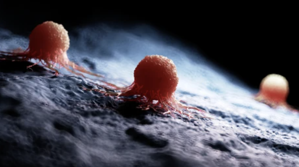
Cancer cells have long been known to divide uncontrollably, but the precise mechanisms that push them into that state have remained only partially understood. A new study from chemists at the Massachusetts Institute of Technology offers a significant piece of that puzzle, revealing how the composition of a cell’s outer membrane can directly influence the behavior of a receptor that governs cell growth.
The research, published in the journal eLife, found that elevated concentrations of negatively charged lipids in the cell membrane can lock a key growth-promoting receptor into a permanently active state, even in the absence of the signals that would normally trigger it. The finding opens a potential new avenue for cancer treatment by targeting the membrane environment rather than the receptor itself.
How the receptor normally works
The epidermal growth factor receptor, known as EGFR, is found on cells that line body surfaces and organs and plays a central role in regulating cell growth. Under normal conditions, the receptor activates only when a specific molecule binds to it, triggering a signaling cascade that tells the cell to divide. When that signal is absent, the receptor remains dormant.
Certain cancers, particularly lung cancer and glioblastoma, are known to overexpress EGFR, which contributes to runaway cell proliferation. What the MIT researchers set out to understand was whether the membrane surrounding the receptor could itself be influencing how the receptor behaves, independent of the usual signaling process.
What happens when lipid levels rise
To investigate, the research team used specially engineered nanodiscs, synthetic structures that mimic the cell membrane and allow scientists to embed full-length receptor proteins for study. Using a technique that measures energy transfer between fluorescent tags attached to different parts of the receptor, the team was able to track precisely how the receptor’s shape changed under different membrane conditions.
Under normal circumstances, roughly 15 percent of the cell membrane consists of negatively charged lipids. The researchers found that membranes containing between 15 and 30 percent negatively charged lipids behaved typically. But when that concentration reached 60 percent, something significant happened. The EGFR receptor locked into its active conformation permanently, continuously signaling the cell to grow regardless of whether any activating molecule was present.
Many cancer cells show exactly this kind of elevated lipid profile. The research suggests that this membrane environment may be one of the mechanisms explaining why those cells are able to sustain unchecked proliferation over time.
The role of cholesterol
The team also examined how cholesterol levels in the membrane affect receptor behavior. When nanodiscs were created with higher cholesterol concentrations, the membranes became more rigid. That increased rigidity had a suppressive effect on EGFR signaling, essentially dampening the receptor’s activity. The finding suggests that membrane stiffness and lipid composition work in tandem to regulate how growth signals are processed at the cellular level.
A new target for treatment
The implications for cancer therapy are meaningful. If negatively charged lipids are driving EGFR into a permanent active state, then neutralizing that charge could offer a way to dial down the proliferative signal without directly targeting the receptor protein itself. That represents a fundamentally different approach from most existing therapies, which focus on blocking the receptor or the molecules that bind to it.
The research was funded by the National Institutes of Health and MIT’s Department of Chemistry. Researchers say further work is needed to explore whether this mechanism can be therapeutically exploited and whether similar lipid-driven effects occur across other membrane receptors involved in cancer progression.