Glioblastoma carries a reputation that few other diagnoses match, and for good reason. The World Health Organization classifies it as a grade 4 astrocytoma, the most aggressive label the agency assigns to tumors that begin in the brain’s star shaped support cells, known as astrocytes. What makes glioblastoma especially difficult to treat is its variety. A single tumor often contains several different types of cells, each behaving differently and responding to treatment in its own way.
Where the tumor sits changes everything
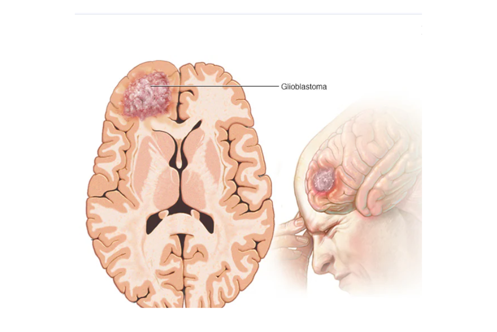
A glioblastoma’s location inside the skull shapes both its symptoms and the treatment options available. As the tumor grows, it presses on and damages healthy tissue, and the effects depend heavily on which part of the brain bears the brunt. A tumor in the frontal lobe can alter someone’s personality, loosen their inhibitions, and make planning daily tasks difficult, sometimes alongside weakness on one side of the body. Involvement of the parietal lobe can bring trouble with speech, writing or math, and a loss of spatial orientation. Tumors in the temporal lobe tend to affect memory and language, while those in the occipital lobe often bring on visual disturbances or partial loss of sight. A tumor pressing on the cerebellum can throw off balance and coordination, and if it reaches the brain stem or spinal cord, it can interfere with breathing, heart rate and blood pressure, in some cases leading to paralysis.
How doctors grade brain tumors
Pathologists assign tumors a grade from one to four based on how the cells look under a microscope and how quickly they are likely to grow and spread. Tumors graded one or two tend to grow slowly and are less likely to push into nearby tissue, while grade three and four tumors grow faster and spread more readily into the surrounding brain. Glioblastoma sits at grade four, the most aggressive category, meaning its cells look highly abnormal, multiply rapidly and extend well beyond the original tumor site.
This aggressiveness also separates malignant tumors from benign ones. A malignant tumor like glioblastoma can grow without restraint, invade nearby structures and in some cases spread to other parts of the body through the bloodstream or lymphatic system. A benign tumor, by contrast, grows slowly and stays put. It can still cause problems depending on its size and location, but it generally poses far less danger than a malignant growth.
Recognizing the signs of glioblastoma
Symptoms vary depending on the tumor’s size, location and rate of growth, but certain patterns show up often. Headaches are common and tend to be severe and persistent, frequently worse in the morning or after a long stretch of sleep. Seizures are another frequent first sign that brings patients to a doctor, and many people also notice changes in memory, speech or other cognitive functions, along with shifts in personality that family members often notice first. Nausea and vomiting can occur as pressure builds inside the skull, while weakness or numbness, usually on one side of the body, points to the tumor’s effect on motor pathways. Vision changes, including blurred sight or a loss of peripheral vision, round out the warning signs that prompt further evaluation.
What raises the risk for glioblastoma
Scientists have not pinned down a single cause of glioblastoma, but a handful of factors raise the odds. Certain inherited genetic conditions, including Li Fraumeni syndrome and Turcot syndrome, are linked to higher risk. Age plays a role too, with glioblastoma showing up more often in adults over 50, and men are diagnosed slightly more often than women. Previous radiation treatment to the brain, often given years earlier for an unrelated condition, has also been tied to a greater likelihood of developing the disease later on.
The path to a diagnosis
Confirming glioblastoma typically unfolds in stages. A neurological exam comes first, checking reflexes, coordination, vision and other functions that might reveal where in the brain something has gone wrong. Magnetic resonance imaging follows, giving doctors a detailed picture of the tumor’s size and location. The final and most definitive step is a biopsy, in which a sample of tumor tissue is examined under a microscope to confirm cancer cells and assign the tumor its grade.